Knee Muscle Anatomy Mri / knee anatomy | MRI knee coronal anatomy | free cross ... / The journal of musculoskeletal medicine.. Learn about mri anatomy with free interactive flashcards. Want to learn more about it? Radiology imaging medical anatomy human anatomy and physiology anatomy study. This mri knee cross sectional anatomy tool is absolutely free to use. Each anatomical structure was labeled interactively.
They move when you do—when you walk, run, dance, stretch your legs, or make any action you can think of that there are two muscle groups that act on the knee joint: Learn anatomy using a full pacs! Click now to learn more about the bones, muscles, and soft tissues of these regions at leg and knee anatomy: This section of the website will explain large and minute details of sagittal knee cross sectional anatomy. Master leg and knee anatomy using our topic page.
Lower extremity: MRI of Anatomical atlas from www.imaios.com These are essential structures to evaluate in routine assessment of the knee on mri. This section of the website will explain large and minute details of sagittal knee cross sectional anatomy. The muscles of the knee joint are incredibly important. An understanding of normal anatomy and biomechanics of the knee extensor mechanism is necessary to comprehend the imaging of extensor mechanism injuries. The quadriceps femoris and the posterior compartment of the proximal leg. Scroll through the structures to understand the anatomy. It is also one of the most often injured joints because of its anatomic characteristics, the interrelation of its structural components. Radiology imaging medical anatomy human anatomy and physiology anatomy study.
Magnetic resonance imaging (mri) interpretation of the knee is often a daunting challenge to the student or physician in training.
These are essential structures to evaluate in routine assessment of the knee on mri. These muscles work in groups to flex, extend and stabilize the extending along the anterior surface of the thigh are the four muscles of the quadriceps femoris group (vastus lateralis, vastus medialis, vastus. Injuries of the patellofemoral joint. This mri knee cross sectional anatomy tool is absolutely free to use. Master leg and knee anatomy using our topic page. Rubin da, kettering jm, towers jd, britton ca: This mri knee cross sectional anatomy tool is absolutely free to use. Knee anatomy francesc malagelada jordi vega pau golanó the knee is the largest joint in the human body and one of the most complex from a functional point of view. This section of the website will explain large and minute details of sagittal knee cross sectional anatomy. In this second module, we will discuss the anatomy and positioning of the bones, joints, ligaments, muscles, blood vessels, and nerves of the lower extremity. Tips to keep joints healthy. The knee is designed to fulfill a number of functions: This mri knee cross sectional anatomy tool is absolutely free to use.
Knee anatomy francesc malagelada jordi vega pau golanó the knee is the largest joint in the human body and one of the most complex from a functional point of view. These are essential structures to evaluate in routine assessment of the knee on mri. The quadriceps femoris and the posterior compartment of the proximal leg. Injuries of the patellofemoral joint. Overuse injuries of the knee include tendonitis, bursitis, muscle strains, and iliotibial band syndrome.
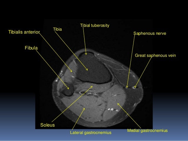
MRI KNEE JOINT ANATOMY from image.slidesharecdn.com The muscles that affect the knee's movement run along the thigh and calf. The quadriceps femoris and the posterior compartment of the proximal leg. Radiology imaging medical anatomy human anatomy and physiology anatomy study. The journal of musculoskeletal medicine. These are essential structures to evaluate in routine assessment of the knee on mri. Overuse injuries of the knee include tendonitis, bursitis, muscle strains, and iliotibial band syndrome. The muscles of the knee joint are incredibly important. Support the body in an upright position without the need for muscles to work.
Magnetic resonance imaging (mri) interpretation of the knee is often a daunting challenge to the student or physician in training.
This mri knee cross sectional anatomy tool is absolutely free to use. Each anatomical structure was labeled interactively. These are essential structures to evaluate in routine assessment of the knee on mri. This mri knee cross sectional anatomy tool is absolutely free to use. Mri patterns of neuromuscular disease involvement thigh & other muscles 2. It is a noninvasive test that can visualize the inner structures of the knee, including the cartilage and ligaments, the surface of the bones, and the muscles and tendons that surround the knee joint. Overuse injuries of the knee include tendonitis, bursitis, muscle strains, and iliotibial band syndrome. Tendons attach the muscles to each other. The knee is designed to fulfill a number of functions: Helps to lower and raise the body. Involved early gray = muscle: Magnetic resonance imaging (mri scan): Injuries of the patellofemoral joint.
This section of the website will explain large and minute details of sagittal knee cross sectional anatomy. Learn anatomy using a full pacs! Support the body in an upright position without the need for muscles to work. This webpage presents the anatomical structures found on knee mri. Tendons attach the muscles to each other.
MRI KNEE JOINT ANATOMY from image.slidesharecdn.com These muscles work in groups to flex, extend and stabilize the extending along the anterior surface of the thigh are the four muscles of the quadriceps femoris group (vastus lateralis, vastus medialis, vastus. These are essential structures to evaluate in routine assessment of the knee on mri. Master leg and knee anatomy using our topic page. Injuries of the patellofemoral joint. Mri for evaluating knee pain in older patients: Learn about mri anatomy with free interactive flashcards. The muscles that affect the knee's movement run along the thigh and calf. General anatomy and musculoskeletal system.
Click now to learn more about the bones, muscles, and soft tissues of these regions at leg and knee anatomy:
Involved early gray = muscle: This section of the website will explain large and minute details of sagittal knee cross sectional anatomy. Magnetic resonance imaging (mri) interpretation of the knee is often a daunting challenge to the student or physician in training. In this second module, we will discuss the anatomy and positioning of the bones, joints, ligaments, muscles, blood vessels, and nerves of the lower extremity. Helps to lower and raise the body. Tendons attach the muscles to each other. Muscles in the posterior compartment of the thigh. Mr imaging of knees having isolated and combined ligament injuries. The muscles of the knee joint are incredibly important. The journal of musculoskeletal medicine. Knee anatomy francesc malagelada jordi vega pau golanó the knee is the largest joint in the human body and one of the most complex from a functional point of view. An understanding of normal anatomy and biomechanics of the knee extensor mechanism is necessary to comprehend the imaging of extensor mechanism injuries. Support the body in an upright position without the need for muscles to work.